
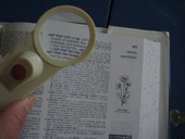


I have Glaucoma. For years, I managed it by taking eye drops regularly seeing an Ophthalmologist every six months to monitor my eyes for changes. Previously, I wrote about my first visit hoping that others could benefit by my experience.
At the end of my last appointment, I asked my ophthalmologist if I could interview someone at the clinic so I could write in more detail about it. He generously arranged for me to spend some time with a Certified Ophthalmic Technician.
Ophthalmologist vs Optometrist
At the appointed time, the Opthalmic Technician greeted me warmly and through our meeting he patiently answered my questions. I began by explaining that before coming to the Eye Care Center, my lifelong eye care consisted of seeing an Optometrist for new glasses every three or four years. I had been surprised by discovering another person in the process.
Ophthalmologists have had technicians and assistants since the early 1970s. These eye-care physicians perform complex surgeries, often on an emergency basis, and they diagnose and treat diseases of the eye that effect our ability to see. They need trained and trusted personnel who can conduct routine examinations, maintain equipment, and collect information from patients.
I learned that Ophthalmologists have a college degree with pre-Med courses, spend four more years getting an MD, then an additional four years studying the eye as a specialty, and after that, studying some part of the eye. In addition to general eye surgeons, there are specialists in the Cornea (front of the eye), the Retina (back of the eye), Glaucoma, Ocular Plastics (blink and lid) and particular diseases of the eye.
Diagnostic Machines and Tests
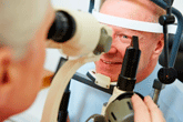
Today, Ophthalmologists have many machines available for painless and non-invasive diagnosis of the eye.
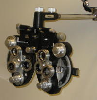
He took me on a tour to see the equipment available at the Eye Care Center. We began with the Phoropter (or Refractor) that has been used since 1938 for identifying the best corrective lenses. He explained that using this machine is an art more than a science because the eye is constantly trying to adjust to what it sees and so can mislead both it's owner and the technician. He told me that modern technology has produced computer electronics that can provide similar results to the work of a trained human technician.
Phoropter or Refractor. A retinoscopy test is done in conjunction with refraction. This test is used to discover how strong glasses need to be.
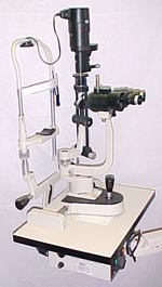
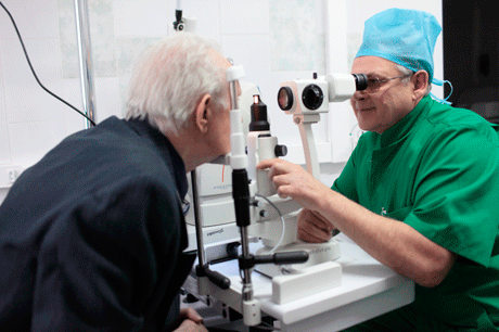
Slit Lamp. A slit lamp uses magnification and light to look at the cornea for defects and irregularities. It also has a Goldman Applination Tonometer to check eye pressures.
Transilluminator.This lighted devise is used to check pupils to see if they are responding equally.
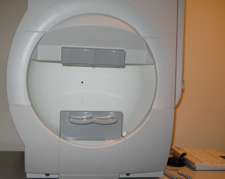
Humphrey Visual Field. Used to analyze peripheral vision both in terms of the field of vision and intensity, or ability to see light. Results are compared to a computer data base of people with similar race and gender characteristics.
Frequency Doubling Technology (FDT). This machine tests the sensitivity of the retinal nerve. The nerve itself is composed of fiber layers. Glaucoma causes a thinning of the layers and therefore a loss of sensitivity to light. Results of FDT tests are also compared to a data base of people of the same age.
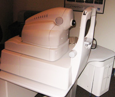
Optical Coherence Tomography (OCT). The OCT measures the retinal nerve fiber layer and also the size and depth of the optic nerve. It is often used to diagnose retinal defects. Test results are compared to a data base or people with the same age, gender and ethnic background.
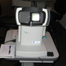
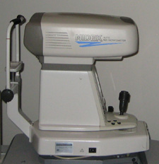
Corneal Topographer. This machine measures the curvature an shape of the cornea. The ophthalmologist is able to identify the strength of the cornea with these test results. In addition, the information is used for cataract surgery preparation and to measure corneal changes.
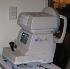
Auto Refractor/Keratometer. This automated machine also measures the cornea for cataract surgery, and to detect changes to the cornea over time.
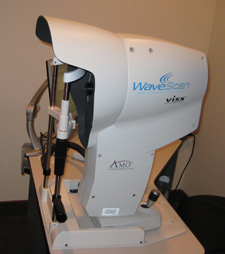
Wave Scan. This machine was designed from technology developed for the Hubble space telescope repair. It identifies all the irregularities in an eye. The information produced is used to program a laser for surgery.
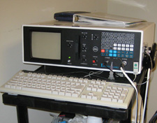
Ultrasound. An ultrasound is used to measure focal length using sound waves to create echoes. These measurements tell the ophthalmologist the thickness of the lens in the eye, the distance from the cornea to the lens, and the distance from the retina from the lens. These measurements are very helpful in cataract surgery.
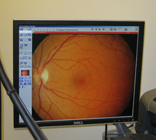
Digital Camera. Images of the retina allow the doctor to watch for changes and make it possible to have more accurate measurement of the optic nerve and other important indicators.